The morphological world of iPSCs — decoding the secrets behind these microscopic lives.
Freshly thawed iPSCs — "shriveled travelers"
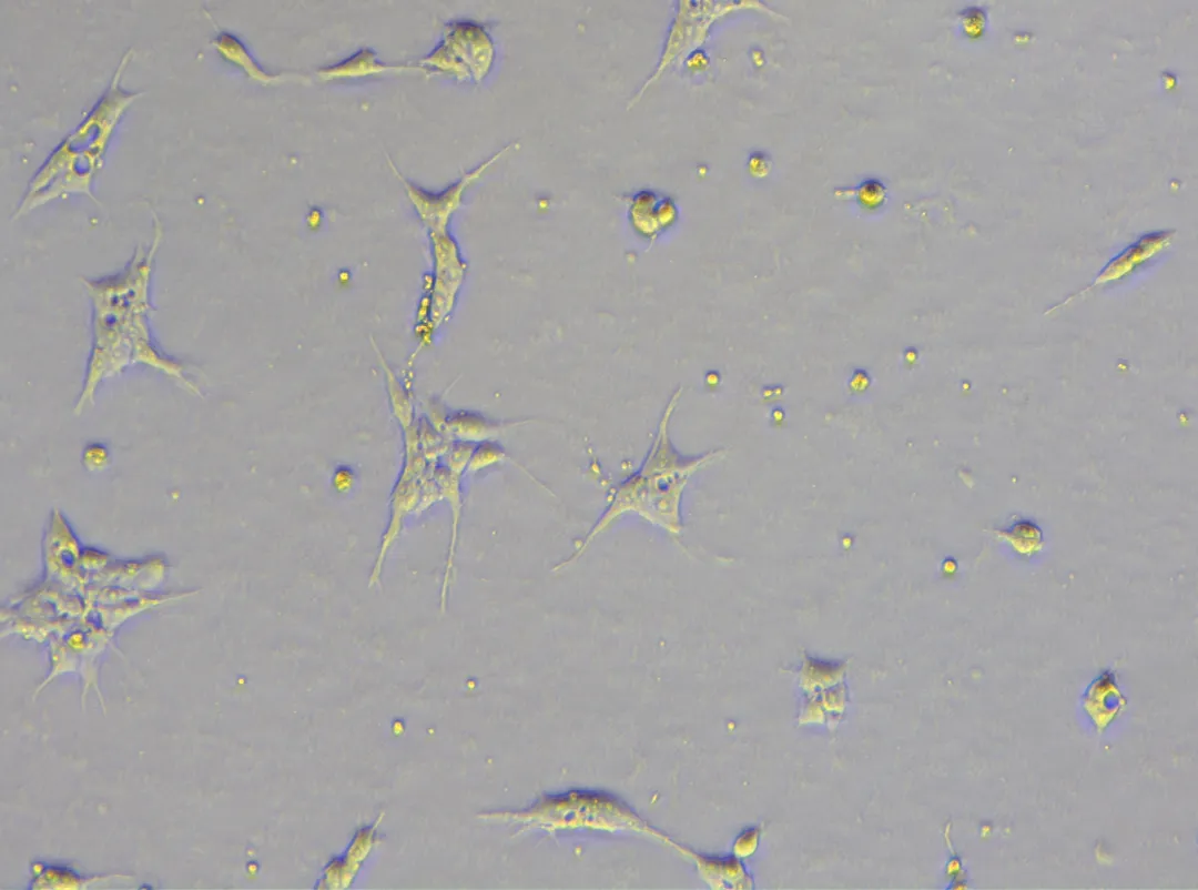
Just after thawing, iPSCs often show poor morphology: cells clump together with indistinct edges, and many dead, floating cells are present. At this stage, we usually add Y27632 (a ROCK inhibitor) to improve cell survival.
However, it is worth noting that Y27632 also temporarily alters cell morphology: cells become elongated, exhibiting unusual "spindle‑like" or "stellate" shapes, and cell‑cell connections become looser. Don't worry — this is the drug in action, protecting cells from apoptosis. It is merely a "temporary protective suit" for the cells.
Acclimated iPSCs — "perfect clonal discs"
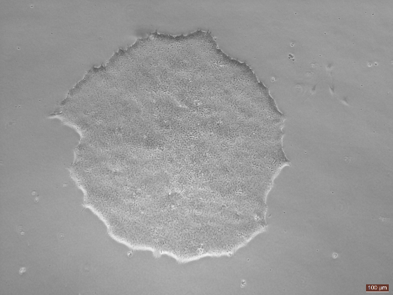
After 2–3 days of recovery and acclimation, iPSCs gradually display their classic morphology:
-Forming dense, well‑defined colonies with sharp borders
-Cells grow as a monolayer, appearing as regular round or oval shapes
-High nuclear‑to‑cytoplasmic ratio with smooth edges
-Cells within the colony are tightly packed, with almost no visible gaps
This is what iPSCs should look like! Uniform and orderly, as if greeting you: "I'm in great condition now!"
The risk before passaging — "little hands of differentiation"
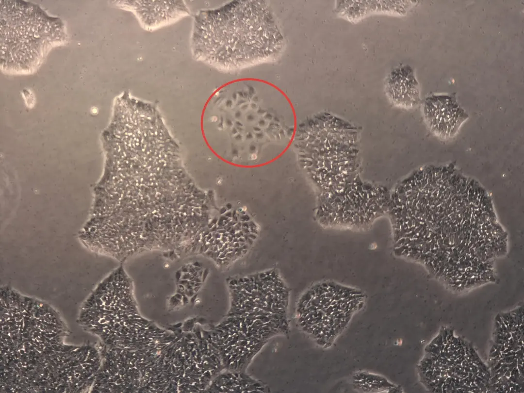
Before passaging, we often observe abnormalities at the edges of some colonies:
-Localized cells become elongated and stretched
-Formation of structures resembling "little hands" or "tentacles"
-Cells in these areas become darker in color with blurred borders
These are early signs of spontaneous differentiation! Once detected, it indicates that you need to passage the cells promptly or remove the differentiated areas; otherwise, the entire colony may become compromised.
Fully differentiated colonies — "punctate outburst"
If a colony becomes fully differentiated, its morphology undergoes a fundamental change:
-The colony loses its original clear borders
-Dense punctate or网状 (reticular) structures appear inside
-Cells may grow in multiple layers, showing明显的隆起 (pronounced bulging)
-Morphological transformation toward neurons, epithelial cells, fibroblasts, etc.
At this point, the colony is no longer suitable for pluripotency studies.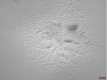
High‑confluence iPSCs — "a continuous landscape of rounded planets"
When cell confluence reaches above 80%, colonies come into contact and merge into a continuous layer:
-Cells remain plump and rounded in morphology
-Borders between colonies disappear, forming a neat monolayer
-The overall appearance resembles a "cellular ocean" — uniform and dense
This is the ideal time for passaging — the cells are in optimal condition.
✨ Morphology Tips:
-
Y27632 temporarily alters cell morphology and should be removed by medium change 24–48 hours after thawing
-
"Little hand"‑like structures are early signs of differentiation and need to be addressed promptly
-
The ideal iPSC morphology: sharp borders, dense and plump, high nuclear‑to‑cytoplasmic ratio
-
Regular observation and timely passaging are key to maintaining iPSC pluripotency
Every observation under the microscope is a direct conversation with your cells. Understanding their morphological language allows you to better master iPSC culture and differentiation.
We hope this simple morphology guide helps you become more proficient on your stem cell culture journey!

 Building 28, No. 18, Lane 99, Chun Guang Road, Minhang District, Shanghai, China
 +86-400-8228-027
 market@somaxbio.com


 Home
Home
