Observe cell morphology under a microscope, and record cell density, adhesion status, dead debris, cell clumps, and whether photographs need to be retained for documentation.
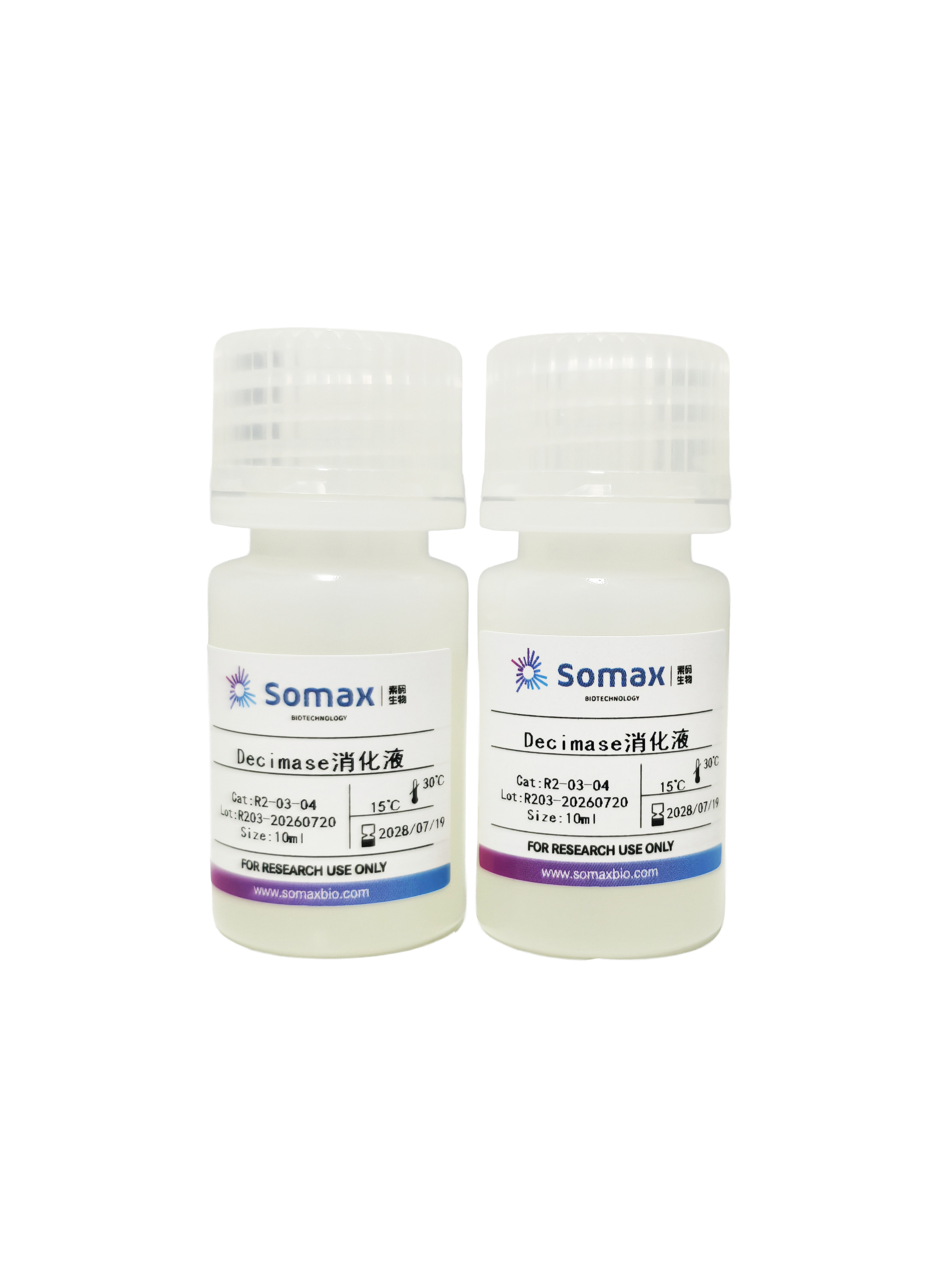
Prepare Decimase Dissociation Solution. Verify the product name, concentration, lot number, expiry date and opening status.
Prepare calcium‑ and magnesium‑free PBS, RPMI 1640 or the corresponding cell maintenance medium for washing, diluting dissociation enzyme and harvesting cells.
It is recommended to pre-warm PBS, RPMI 1640 or the corresponding medium to 37°C.
If cells tend to aggregate after dissociation, it is recommended to prepare cut-tip 1 mL pipette tips in advance for gentle pipetting and clump dissociation in subsequent steps.
At least two 15 mL centrifuge tubes are recommended per sample: one for primary harvesting and one for transfer after secondary dissociation following centrifugation.
Aspirate the spent medium, wash cells once gently with pre-warmed PBS and remove PBS completely to avoid dilution of Decimase Dissociation Solution by residual liquid.
Add 1 mL of Decimase Dissociation Solution per well to cover the cell monolayer. Rock the plate gently for even distribution of the enzyme solution, place it in a 37°C incubator to start dissociation and record the start time. Examine cell morphology under a microscope every 10 minutes thereafter (the suggested dissociation time is 15–30 minutes and may be adjusted according to cell status).
Note: The dissociation endpoint can be judged by the following signs: the cell monolayer begins to loosen overall, the peripheral regions can be lifted by gentle pipetting, most cells change from spread morphology to round or semi-detached status, and cells can detach from the culture surface upon gentle pipetting. If these criteria are not met, continue incubation and inspect at 10-minute intervals. Repeated vigorous pipetting is not recommended when dissociation is incomplete, as it may cause localized cell damage or heterogeneous clump formation.
When the dissociation endpoint is reached, add 2 mL of pre-warmed RPMI 1640 supplemented with 10 μM Y27632 or the corresponding cell maintenance medium containing 10 μM Y27632 to quench dissociation. Use a cut-tip 1 mL pipette tip to rinse the culture surface gently to facilitate cell detachment. If substantial clumps remain, continue gentle pipetting with the cut-tip 1 mL pipette tip; avoid generating excessive foam and prevent repeated scraping of the cell monolayer with the pipette tip. Transfer the cell suspension into a 15 mL centrifuge tube. Rinse the culture vessel once with medium from the centrifuge tube to maximize cell recovery.
5. Centrifuge at 200 g for 5 minutes. Carefully aspirate the supernatant after centrifugation without disturbing the pellet. Cell clumps may be dispersed by gentle pipetting using a cut-tip 1 mL pipette tip.
Note: Some cells tend to re-aggregate after centrifugation. For cell types prone to clumping such as cardiomyocytes, a second round of gentle dissociation is recommended. Procedure: Observe the cell pellet and clumps after centrifugation, add an appropriate volume of pre-warmed medium or medium required for subsequent steps. Gently pipette using a cut-tip 1 mL pipette tip, transfer the well-dispersed homogeneous cell suspension into a new 15 mL centrifuge tube. Continue gentle pipetting on the remaining large clumps in the original tube. Stop pipetting when only a small amount of filamentous or hard-to-disperse aggregates remain to avoid excessive mechanical damage.
Gently resuspend cells with Cardiomyocyte Seeding Medium (R3-01-015). Take an aliquot for cell counting and viability assay. It is recommended to record total cell count, viable cell count, viability, residual clumps and filamentous debris, then proceed to passaging, re-seeding, cryopreservation or subsequent functional assays according to experimental objectives.
Precautions
(1) Verify the lot number, expiry date and opening status of Decimase Dissociation Solution before use.
(2) Remove PBS completely after washing to prevent dilution of dissociation enzyme by residual PBS.
(3) Dissociation time should be adjusted according to cell type, adhesion strength, cell density and culture vessel.
(4) Vigorous pipetting in the early stage of dissociation is not recommended for sensitive or tightly adherent cells.
(5) If cells tend to aggregate, use cut pipette tips for gentle pipetting instead of harsh mechanical force for forced dispersion.
(6) If cell clumps grow larger after centrifugation, perform gentle dispersion prior to counting or downstream processing.
(7) Hard-to-disperse filamentous debris may be recorded as process loss. Excessive pipetting or filtration for full recovery is not advised.
Scope of Application
This product is for research use only and is not intended for clinical therapy or other applications.




 Home

 Building 28, No. 18, Lane 99, Chun Guang Road, Minhang District, Shanghai, China
 +86-400-8228-027
 market@somaxbio.com